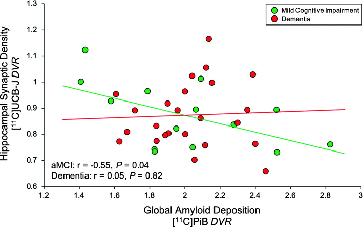
Fig. 2.
Correlation of global Aβ deposition and hippocampal SV2A in aMCI and dementia due to AD. Scatter plot with best-fit lines depicts a significant inverse association between global Aβ deposition and hippocampal SV2A in participants with aMCI (green) but not with dementia (red). Correlation coefficients were calculated from separate univariate linear regression analyses in each group with associated two-tailed P values, without correction for multiple comparisons. Global Aβ deposition was calculated by averaging values of [11C] PiB DVR from the bilateral prefrontal, lateral temporal, posterior cingulate/precuneus, and lateral parietal ROIs, weighted by volume. Green circles denote DVR values for aMCI participants, while red circles denote DVR values for participants with dementia. Abbreviations: DVR, distribution volume ratio using a whole cerebellum reference region; aMCI: amnestic mild cognitive impairment; SV2A, synaptic vesicle glycoprotein 2A; PiB, Pittsburgh Compound B

