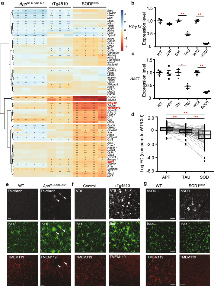
Fig. 2.
Decreases in gene expression linked to homeostatic microglia in mouse models of neurodegenerative diseases. Expression of homeostatic microglial genes analyzed by RNA sequencing (RNA-seq) and quantitative PCR (WT: n = 4 and AppNL-G-F/NL-G-F: n = 4; Ctrl: n = 3 and rTg4510: n = 3; WT: n = 4 and SOD1G93A: n = 4). WT, wild-type; Ctrl, control. a A heat map of homeostatic microglial genes in isolated-microglia of the mouse models of neurodegenerative diseases. Colors of the individual cells denote relative expression levels of the neurodegenerative models. +: q < 0.05, ++: q < 0.001. b, c Quantitative PCR analysis to determine the expression levels of P2ry12 and Sall1 mRNA in isolated-microglia of each mouse model. b P2ry12 expression level (AppNL-G-F/NL-G-F: FC = −1.14, p = 0.2275; rTg4510: FC = −2.07, p = 0.00285; SOD1G93A: FC = −7.35, p = 1.74E−06). c Sall1 expression level (AppNL-G-F/NL-G-F: FC = −1.02, p = 1; rTg4510: FC = −2.21, p = 0.0420; SOD1G93A: FC = −4.04, p = 6.71E−05). Data are represented as the mean ± SEM. *p < 0.05, **p < 0.001. Bonferroni-corrected Student’s t test. d Log2FC values against WT/Ctrl for 68 homeostatic microglial genes in the isolated microglia from the cortex of APPNL-G-F/NL-G-F and rTg4510 mice, and spinal cord of SOD1G93A mice. AppNL-G-F/NL-G-F (median log2FC = 0.227), rTg4510 (median log2FC = −0.0504, adj. p = 7.33E−07), and SOD1G93A (median log2FC = −0.633, adj. p = 1.61E−11) mice. Data are represented as the median with 5th and 95th percentile. *p < 0.05, **p < 0.001. Wilcoxon’s signed rank test, followed by a multiple testing correction using the Bonferroni–Holm method. e–g Representative immunofluorescent images demonstrating amyloid β (Aβ, thioflavin), Tau (AT8) or hSOD1 (white), Iba1 (green), and TMEM119 (red) in cortex of e WT and AppNL-G-F/NL-G-F mouse, f Ctrl and rTg4510 mouse, and g spinal cord of WT and SOD1G93A mouse. Arrowheads indicate Aβ and Aβ-associated microglia. Scale bars: 20 µm (e, f) and 50 µm (g)

