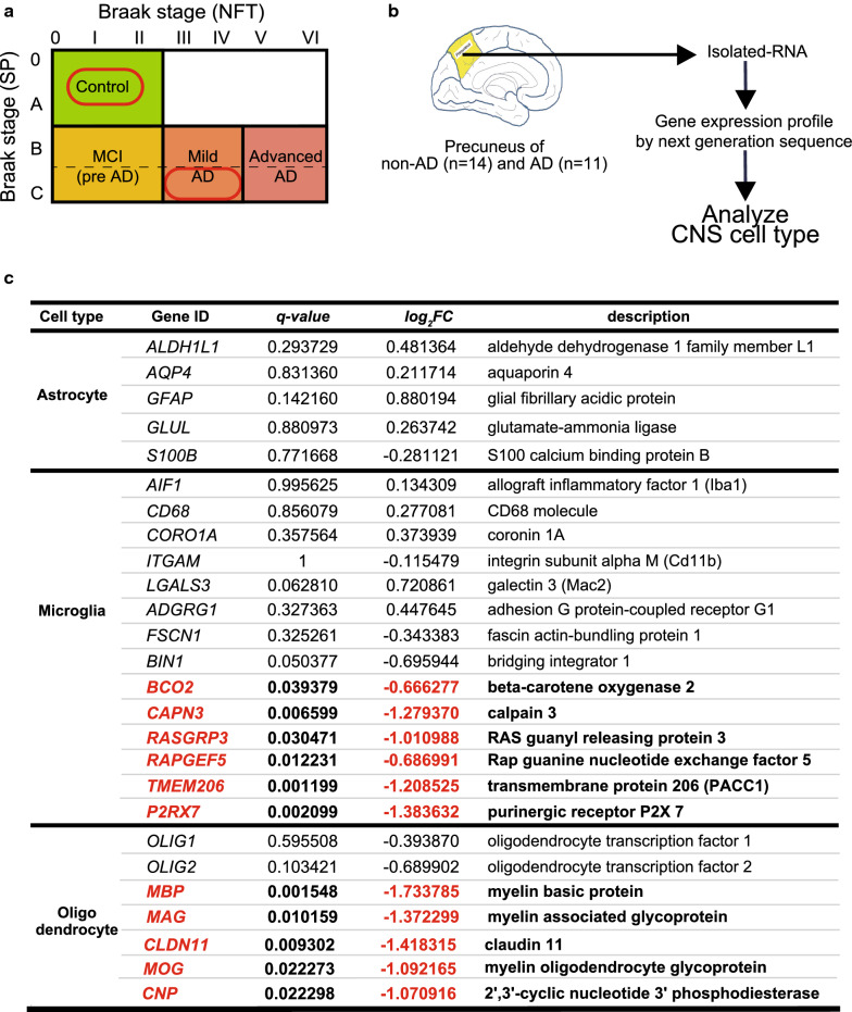
Fig. 4.
RNA sequencing reveals altered gene expression of each CNS cell-type in human precuneus with Alzheimer’s disease (AD) pathology. a Human brain samples were selected for analysis based on the Braak staing as follows: control brain (non-AD) defined as Braak stage (senile plaque: SP): 0–A, Braak stage (neurofibrillary tangle: NFT): 0–II; and AD brain defined as Braak stage (SP): C and Braak stage (NFT): III–IV. b Schematic overview of the gene expression analysis of the CNS cell-type markers in the precuneus of non-AD (n = 14) and AD (n = 11). c Expression of representative genes enriched in astrocytes, microglia, and oligodendrocytes in precuneus of AD brain with fold change. Downregulated genes (statistically significant, q < 0.05) are shown in red and bold

