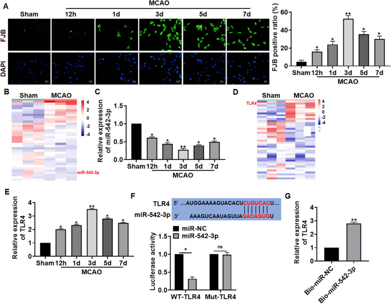
Fig. 1.
The expression of miR-542-3p and TLR4 in brain tissues during cerebral infarction. Mice suffered from sham or MCAO operation with different time points (12 h, 1 days, 3 days, 5 days, and 7 days). a FJB staining was used to detect the degenerating neurons in brain tissues. b MicroRNA expression profiles in sham and MCAO (3 days) mice. Scale bar 20 μm. c qRT-PCR analyzed the expression of miR-542-3p in brain tissues of different groups. d MiRNA expression profiles in sham and MCAO (3d) mice. e TLR4 level in brain tissues of different groups was examined. f The pairing bases between TLR4 and miR-542-3p, and luciferase assay was used to determine the binding of TLR4 and miR-542-3p in HEK293 cells. g RIP assay for the binding of TLR4 and miR-542-3p in HA1800 cells. Data are mean ± SD; *P < 0.05, **P < 0.01. All experiments were repeated in triplicate

