Fig. 1.
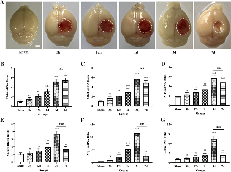
Dynamic changes in mRNA expression of microglial/macrophage M1-like and M2-like phenotypic markers following TBI. a Representative photographs of whole brains in the sham and different traumatic brain injury (TBI) groups (at 3 h, 12 h, 1 day, 3 days, and 7 days post-insult, respectively). Scale bar = 1 mm. Quantitative reverse transcription-polymerase chain reaction (RT-PCR) was used to assess the mRNA expression levels of microglial/macrophage M1-like and M2-like phenotypic markers in the injured cortex at 3 h, 12 h, 1 day, 3 days, and 7 days after TBI or the equivalent area of the sham-operated brains. b–d Expressions of mRNA of M1-like phenotypic markers, including CD16 (b), CD32 (c), and iNOS (d), were gradually increased over time from 12 h onward and remained elevated for at least 7 days after injury. e–g Expressions of mRNA of M2-like phenotypic markers, including CD206 (e), Arg-1 (f), and IL-10 (g), were significantly upregulated at 1 day, 12 h, and 1 day after TBI, respectively, and all peaked around day 3 post-injury, then declined dramatically on day 7, even though they did not return to baseline levels. In b–g, data are expressed as fold change. n = 6 mice per group. ns, nonsignificant, p > 0.05; *, p < 0.05; **, p < 0.01; ***, p < 0.001; versus sham-operated controls; NS, nonsignificant, p > 0.05; ###, p < 0.001; versus the TBI group on day 3. One-way ANOVA followed by Bonferroni’s post hoc tests

