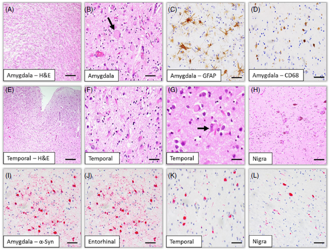
Fig 2.

Histological (A, B, E-H) and immunohistochemical (C, D, I-L) findings of the brain. Histological findings show severe neuronal loss and spongiform changes associated with astrocytosis and microgliosis involving the temporal lobe including the amygdala (A, C–F). Ballooned neurons and eosinophilic ytoplasmic inclusions are noted (B, G). Severe degeneration is observed in the substantia nigra (H). Phosphorylated α-synuclein immunohistochemistry reveals numerous Lewy bodies and Lewy neurites in the amygdala (I), the entorhinal (J) and temporal (K) cortices, and the substantial nigra (L). Immunohistochemistry for GFAP (C) and (CD68 (D) reveals reactive astrocytosis and microgliosis, respectively. Scale bars: 200 μm (A), 200 μm (E), 50 μm (B–D, F–L).
