
Fig. 4 |. A role for the mechanics of stratified cells in tumour invasion.
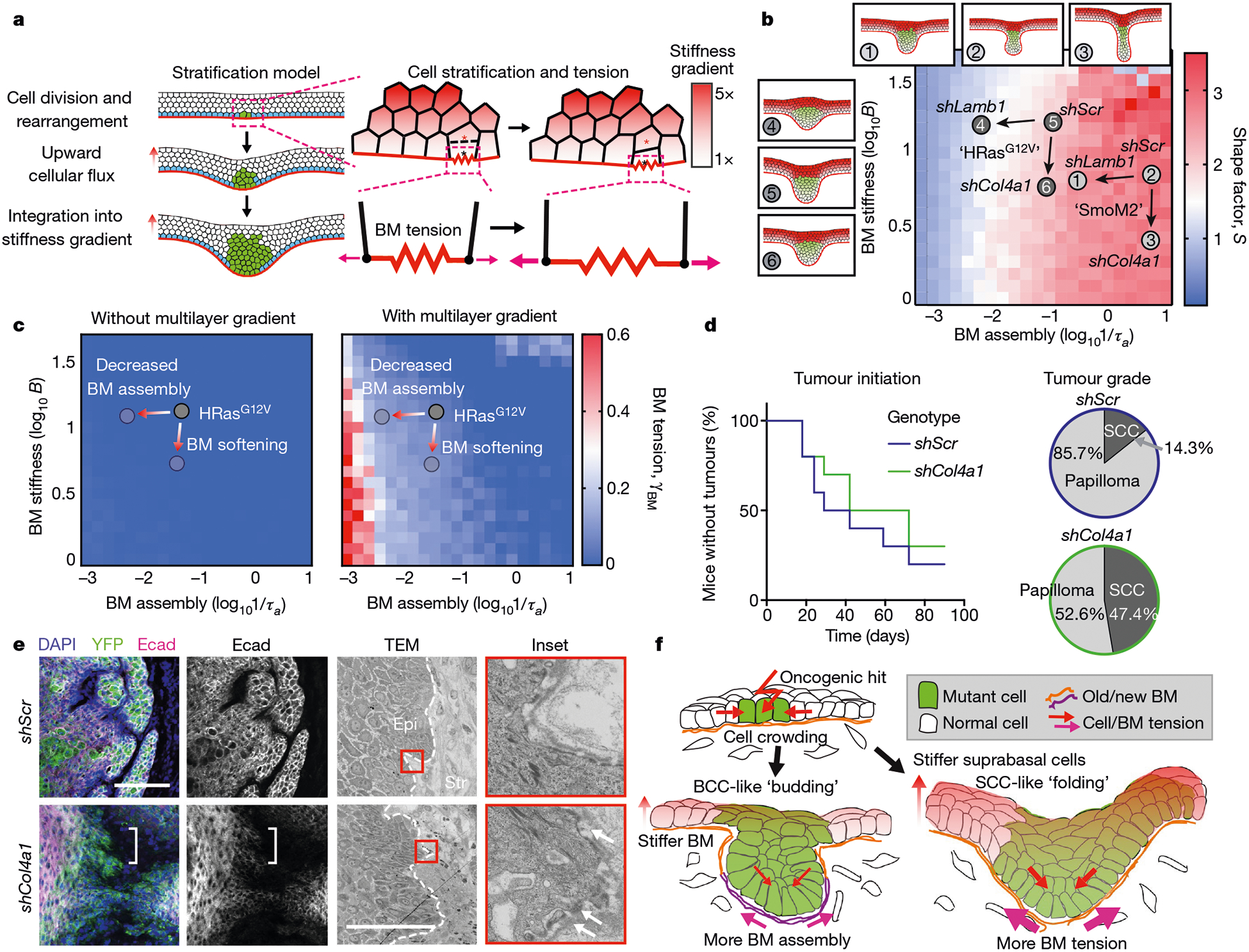
a, Multilayered epithelial vertex model. Tumour cells move upwards into suprabasal layers in a manner that depends on junctional tension and division orientation, while lateral cell tension increases as a function of vertical position. Red and black asterisks indicate a pair of dividing cells. b, Comparison of S values between experimental data and multilayer vertex model simulations that include a suprabasal stiffness gradient. S values are indicated by heatmap. Tissue architecture examples for experimental parameter values of shScr, shLamb1 and shCol4a1 are shown. Arrows denote changes in basement-membrane properties due to shRNAs. c, Changes in BM tension resulting from the multilayer stiffness gradient. Extensile tensions acting on the BM were calculated in the absence (left) and presence (right) of this gradient. d, Impact of decreasing BM stiffness (shCol4a1) on the percentage of mice bearing HRasG12V tumours over time. Papillomas and SCCs were distinguished by tumour pathology upon completion of the experiment (shScr, n = 10 mice; shCol4a1, n = 10 mice). e, Immunofluorescence and ultrastructure imaging of shCol4a1 versus shScr tumours from age-matched littermates. Regions with Ecad downregulation (left, brackets) and BM discontinuities (right, arrows) are indicated. Scale bars, 50 μm. f, Summary of the mechanical forces that affect tumour architecture and invasion.