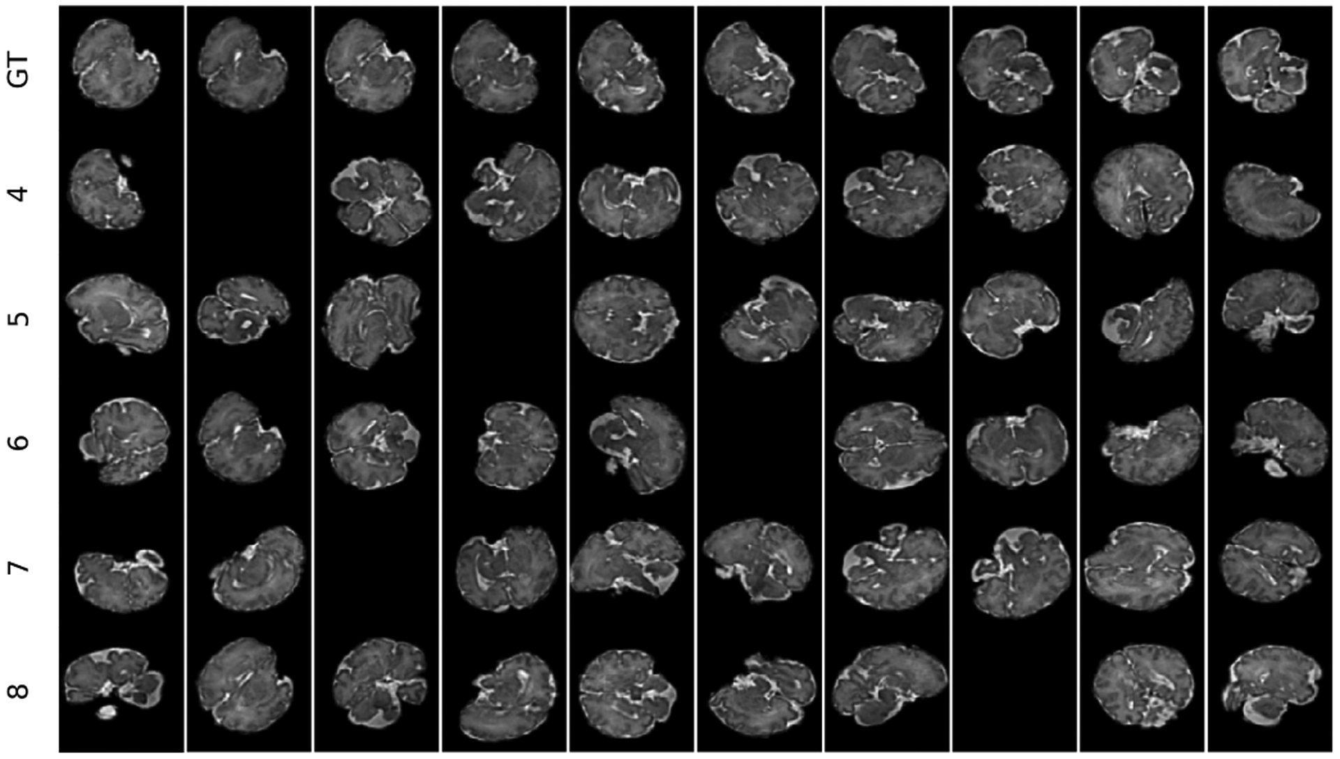
Figure 4:

A demo of five sequences of 10 timesteps each generated with different speeds of motion (corresponding to the number of spline control points from 4 to 8) from the 3D reconstructed fetal brain MRI scan of GA 35 weeks (shown at the top row). Randomly masked slices indicate slices corrupted by intra-slice motion.
