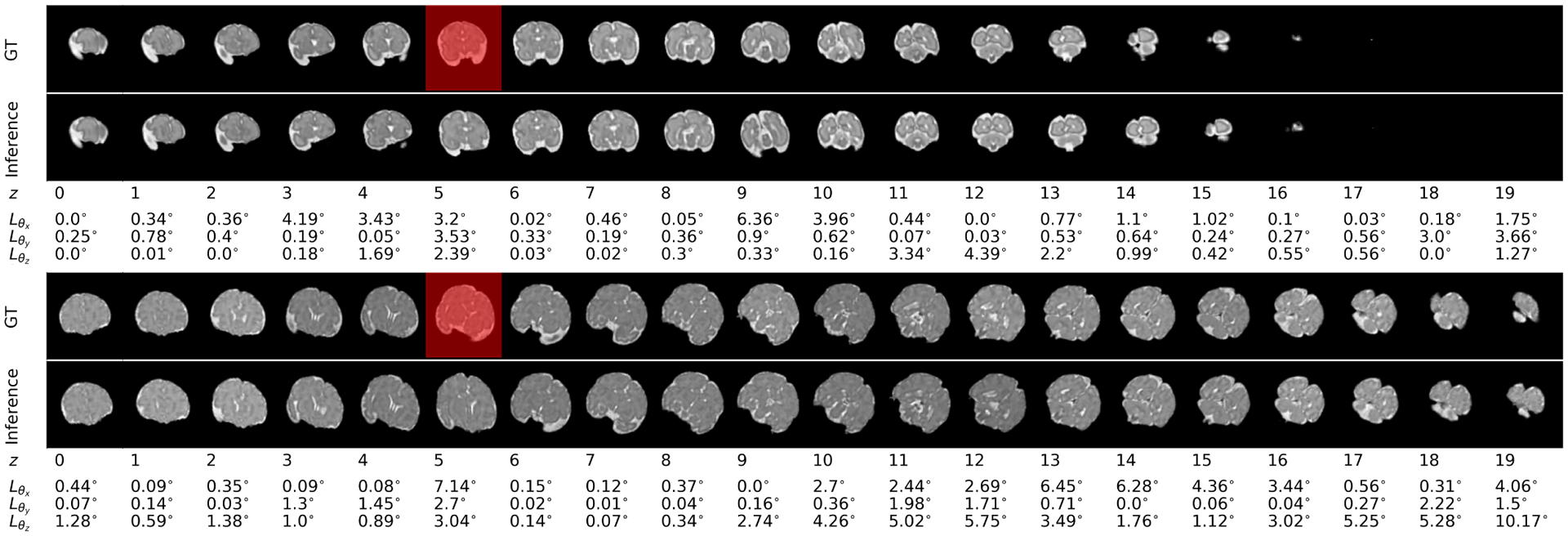
Figure 5:

Inference (i.e. estimation for the first 10 timesteps and prediction for the rest of the 10 timesteps) in the bottom rows has been compared to the ground truth sequence in the top rows for scans of two fetuses: the first figure is a scan of a 28-week, and the second figure is a scan of a 36-week GA fetus from the test set. Errors based on the MSE loss (Section II-F) have been shown underneath each timestep. In these figures the slices shown with red masks were masked in the input sequence. It can be seen that the estimated slices (in the bottom rows) corresponding to the masked slices, showed relatively larger error, but the masked slices did not have a major effect on predictions. Slight increase in prediction error with prediction time horizon was seen in the test sequence, but the predictions were overall accurate.
