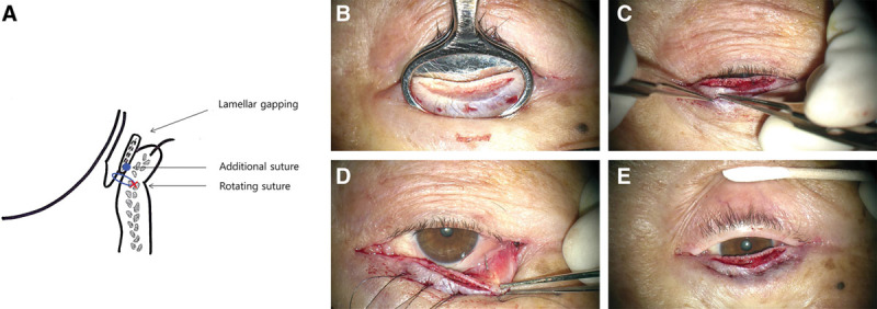
Fig. 1.

A, Schematic view of the eyelid section after the two-step procedure. B, With chalazion forceps, a split incision is made along the gray line. When anterior lamella was separated sufficiently, there was lamella gapping. C–D, Nylon suture passed from skin to palpebral conjunctiva through the inferior border of tarsal plate: everting suture. E, Adjustment of the position of the notes to make buried sutures.
