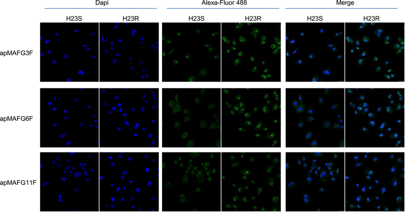
Fig 4.
MAFG localization pattern in H23S and H23R cells measured by aptacytochemistry. Cells were incubated using digoxigenin-labeled aptamers apMAFG3F, apMAFG6F, and apMAFG11F as primary recognition molecule and Alexafluor 488-conjugated anti-digoxigenin as secondary antibody. Confocal microscopy images corresponding to the staining of nuclei with Dapi (blue), aptamers (green) and merge are shown. Bar = 25 μm. MAFG, musculoaponeurotic fibrosarcoma oncogene family, protein G.

