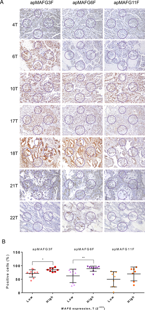
Fig 5.
Analysis of MAFG localization pattern in paraffin samples of patients with known levels of MAFG by aptahistochemistry. Samples were incubated with digoxigenin-labeled aptamers apMAFG3F, apMAFG6F, and apMAFG11F as primary recognition molecules, and afterward with anti-digoxigenin antibodies. Staining was performed using DAB staining solution. (A) Representative aptahistochemistry pictures showing the areas of analysis in NSCLC tumor samples; (B) T test comparison between low and high MAFG mRNA levels (2−ΔCt) and the % mean ratio of positive cells analyzed in the areas defined on (A). Positive cells were counted using the Cell Counter on ImageJ software. NSCLC: non-small cell lung cancer. MAFG, musculoaponeurotic fibrosarcoma oncogene family, protein G

