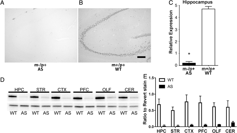
Figure 2.
Characterization of Ube3a deletion rat CNS. (A) Representative anti-E6AP immunohistochemical images of the hippocampus of AS and WT brains showing little to no E6AP detected in the AS rat brain. There were also no obvious gross anatomical aberrations between groups. Scale bar = 200 μm. (B) Relative anti-E6AP staining for the rat hippocampus shows a significant reduction in E6AP staining (AS n = 10 (5M, 5F); WT n = 10 (5M, 5F); t(18) = 17.88, P < 0.05). (C) Western blot with anti-E6AP antibody demonstrating a significant and near-complete reduction of Ube3a within various brain regions. (D) Quantitation of western blot panel C (relative to total protein from Revert staining). HPC, hippocampus; STR, striatum; PFC, prefrontal cortex; CX, rest of cortex; CER, cerebellum; OLF, olfactory bulbs.

