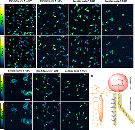
Fig. 5. Polarization-sensitive SRS imaging of AmB-treated various C. auris strains at 1556 cm−1.

(A1 to G1) SRS images of AmB-treated C. auris 1_MGH, C. auris 1_CDC, C. auris 2_CDC, C. auris 3_CDC, C. auris 4_CDC, C. auris 7_CDC, and C. auris 9_CDC at 1556 cm−1 with the laser polarization in the horizontal direction. (A2 to G2) SRS images of the same strains on the same field of view but with laser polarization in the vertical direction. (H) Orientation of ─CH2 in phospholipid backbone versus that of AmB. AmB: 3.2 μg/ml, 1-hour treatment at 30°C. Pixel dwell time: 50 μs. Scale bar, 10 um. Pump, 895 nm; Stokes, 1040 nm. Laser polarization direction is indicated by red arrows.
