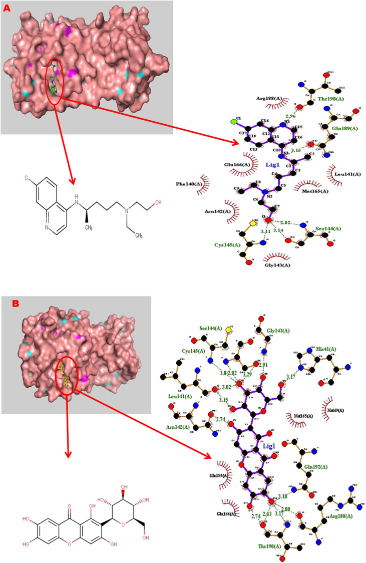
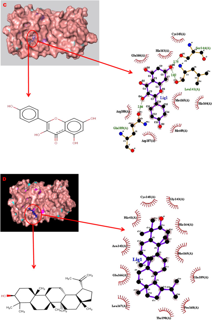
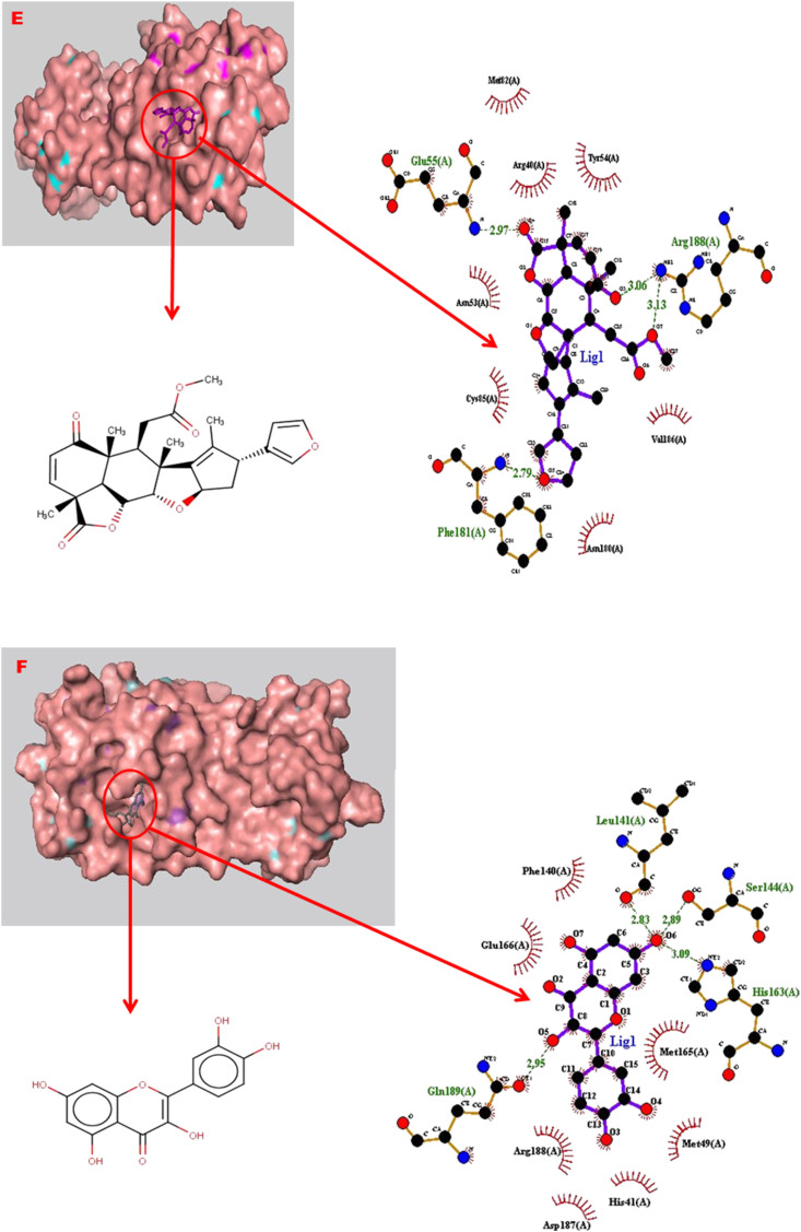
Figure 4.
The binding configuration of ligands showing their poses and interactions in the binding site of the main protease of SARS-CoV-2. (a) Hydroxychloroquine, (b) mangiferin, (c) kaempferol, (d) lupeol, (e) nimbolide, and (f) quercetin. The interaction analysis shows hydrogen bonds (dashed green lines) and hydrophobic interactions (curved red lines) as ligands (purple) interact with the amino acid residues in the active site of Mpro.


