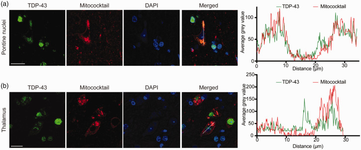
Figure 5.
Colocalization between cytoplasmic TDP-43 and mitochondria. (a and b) Confocal microscopic images demonstrated colocalization between cytoplasmic TDP-43 accumulation and mitochondria in neurons in two representative specific brain regions: pontine nuclei (a) and thalamus (b). Right, line-scan analysis along the solid white lines drawn in the merged images on the left. Each scale bar = 20 μm. (A color version of this figure is available in the online journal.)

