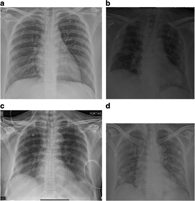
Fig. 1.
Chest X-ray AP portable in 4 different patients showing different lung findings in COVID-19 infection. Technical factors: 70 Kv and 20 mAs. Image a shows normal lungs. Image b shows peripheral consolidation on the right and peripheral lung opacities on the left. Image c shows peripheral ground glass opacification in mid and lower zones. Image d shows typical “batwing” appearance of peripheral consolidation

