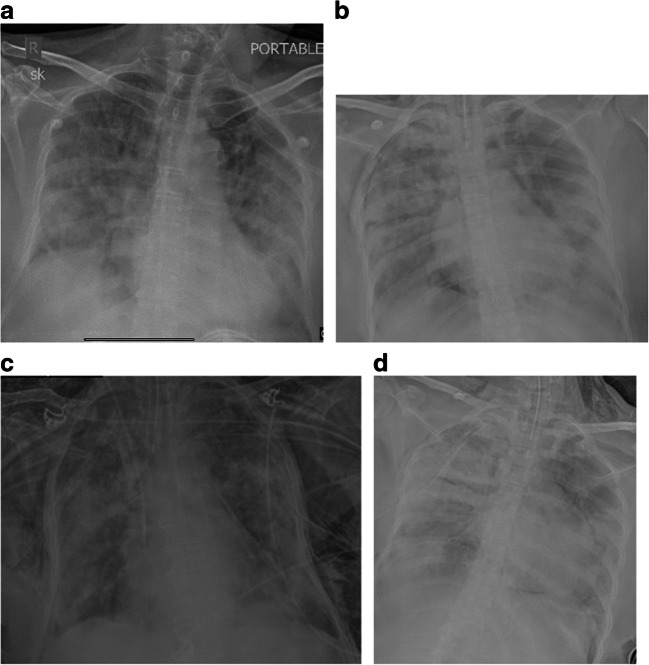
Fig. 2.
Chest X-ray AP portable in 2 patients showing radiological progression. Technical factors: 70kV and 20 mAs. Images a and b show generalized confluent consolidation of both lungs on a background of ground glass haziness with additional features of pneumothorax and pneumomediastinum. Images c and d show bilateral intercostal drains for pneumothorax and extensive subcutaneous emphysema. An additional feature of pneumomediastinum is also seen in image d

