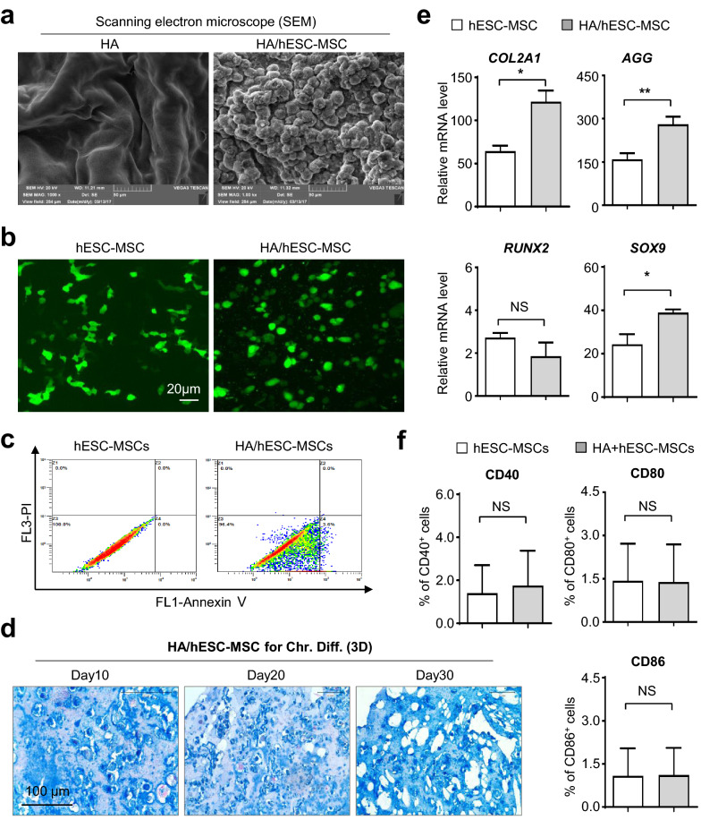
Fig. 6.
HA hydrogels enhanced in vitro chondrogenic differentiation of hESC-MSCs whereas minimally affected cell vitality. a Scanning electron microscope (SEM) analysis of HA hydrogels (HA) and HA/hESC-MSCs hydrogel (HA/hESC-MSCs). b Immunofluorescence analysis of F-ACTIN cytoskeleton of monolayer cultured hESC-MSCs and HA/hESC-MSCs. Scale bar = 20 μm. c FCM analysis of apoptotic population in hESC-MSCs and HA/hESC-MSCs. d The dynamic images of sections of HA/hESC-MSCs-derived chondrocytes with Alcian Blue staining. Scale bar = 100 μm. e qRT-PCR analysis of the chondrogenic-associated genes in the hESC-MSCs and HA/hESC-MSCs groups. f FCM assay of CD40+, CD80+ or CD86+ cells in hESC-MSCs or HA/hESC-MSCs. All data were shown as mean ± SEM (n = 6). *P < 0.05, **P < 0.01; NS, not significant

