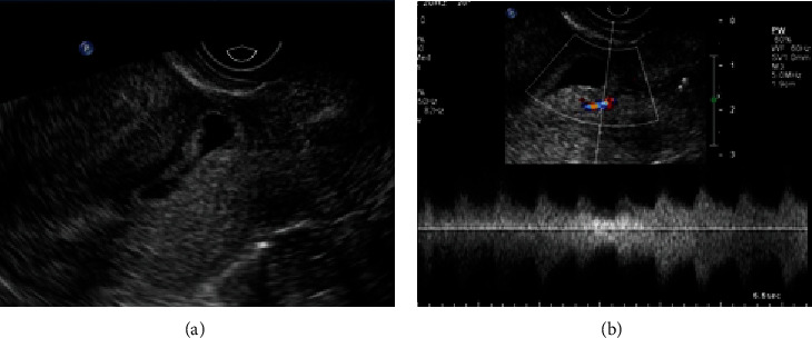
Figure 4.

A 36-year-old female (gravida 4, para 1) presented with amenorrhea for 47 days. (a) The sagittal grayscale image showed that the sac was implanted in the lower endometrial cavity, protruding into the scar. (b) The color Doppler image showed the trophoblastic blood flow from the lower posterior uterus. Chorionic villi were not visible inside the scar with a laparoscopy.
