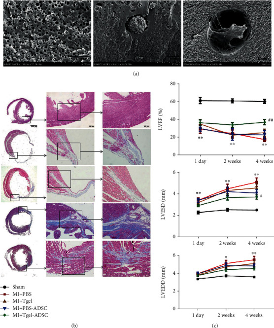
Figure 6.

Scanning electron micrographs of Col-T gel-encapsulated ADSCs 3 days after encapsulation (a). Representative images of Masson trichrome staining of the transverse planes of heart sections (b). LVEF, LVESD, and LVEDD at 1 day, 2 weeks, and 4 weeks after myocardial infarction (c). LVEF: left ventricular ejection fraction; LVESD: left ventricular end-systolic diameter; LVEDD: left ventricular end-diastolic diameter. Adapted from a previous study [144], with permission.
