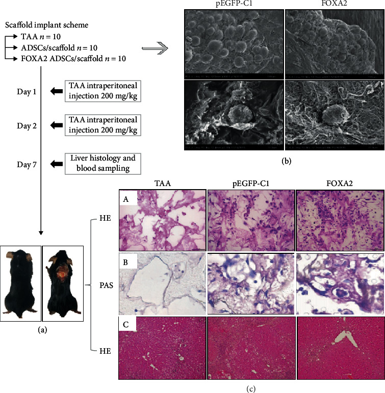
Figure 7.

A schematic representation of the experimental design (a). Scanning electron micrographs of ADSCs in a pEGFP-C1-transfected ADSCs/scaffolds and FOXA2-transfected ADSCs/scaffolds (b). Hematoxylin and eosin (H&E) staining of the necrotic area and retrieved scaffolds (c). TAA: thioacetamide. Adapted from a previous study [158], with permission.
