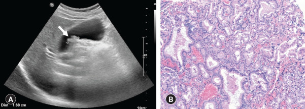
Fig. 3.
Adenomatous gallbladder polyp in a 69-year-old man. (A) Ultrasonography shows a sessile polyp (arrow) with a wide base that was immobile and lacks an acoustic shadow. The polyp measured 16.8 mm in maximal diameter. (B) Microscopically, the adenoma is composed of closely packed, pyloric-type glands lined by mucin-containing cuboidal cells (hematoxylin and eosin stain, x100).

