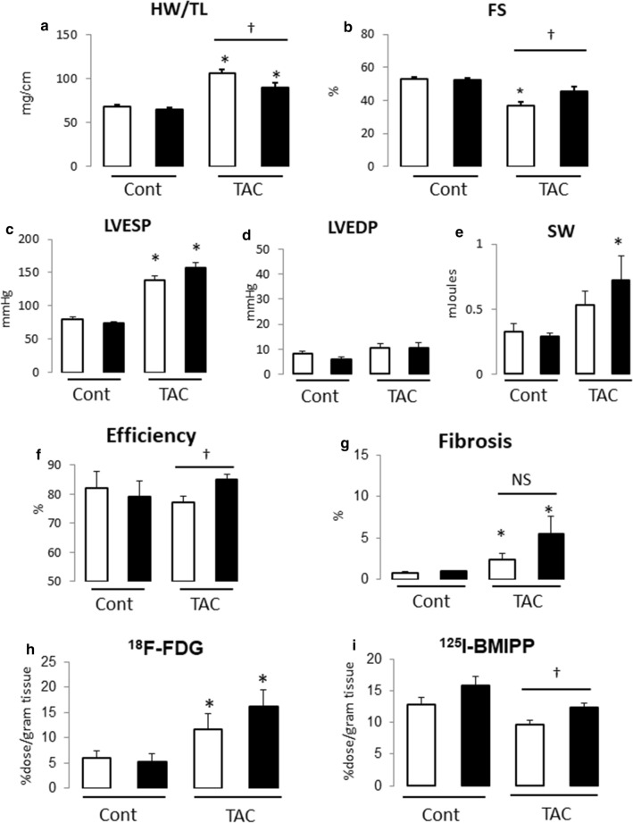
Fig. 1.
Effect of vilda treatment on cardiac hypertrophy and dysfunction in pressure-overloaded non-diabetic mouse heart. a Heart weight/tibia length ratio (HW/TL). b Fractional shortening (FS) measured by echocardiography. Open bar, vehicle-treated; closed bar, vilda-treated (a, b; Cont with vehicle, N = 6; Cont with vilda, N = 6; TAC with vehicle, N = 9; TAC with Vilda, N = 10). c–f Conductance catheter measurements (N = 5–7 of each group). Open bar, vehicle-treated; closed bar, vilda-treated. LVESP, left-ventricular end-systolic pressure (c); LVEDP, left-ventricular end-diastolic pressure (d); SW, stroke work (e); efficiency estimated by SW/pressure–volume area (f). g Myocardial fibrosis area estimated by Masson’s trichrome staining. h, i Glucose uptake and free fatty acid (FFA) uptake in hypertrophied heart and vilda’s effect. Glucose uptake was estimated by 18F-FDG (2-fluorodeoxyglucose) uptake in heart (h). FFA uptake was estimated by 125I-BMIPP uptake in heart (i). Statistical analysis was performed unpaired two-tailed Student’s t test with Welch’s correction. *P < 0.05 vs control. †P < 0.05 vs vehicle-treated group

