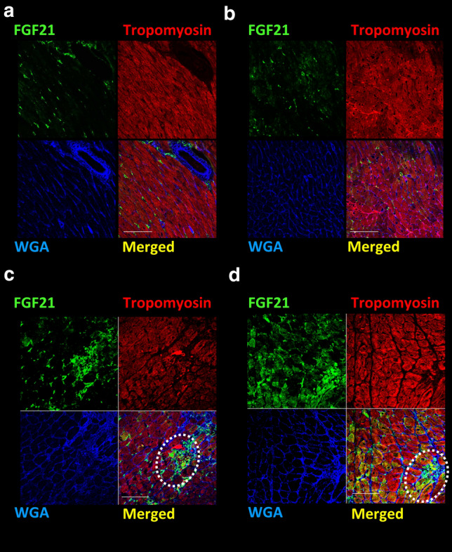
Fig. 4.

Fluorescent immunostaining of heart sections from 3-week TAC mice. Representative fluorescent immunostaining images. Vehicle-treated sham-operated (a), vilda-treated sham-operated (b). Vehicle-treated TAC-operated (c) and vilda-treated TAC-operated (d). FGF21 (green), Tropomyosin (myocytes) (red), and WGA staining (membrane/extracellular matrix) (blue). Dotted circles indicate non-myocyte positive staining for FGF21. Scale bars: 50 µm
