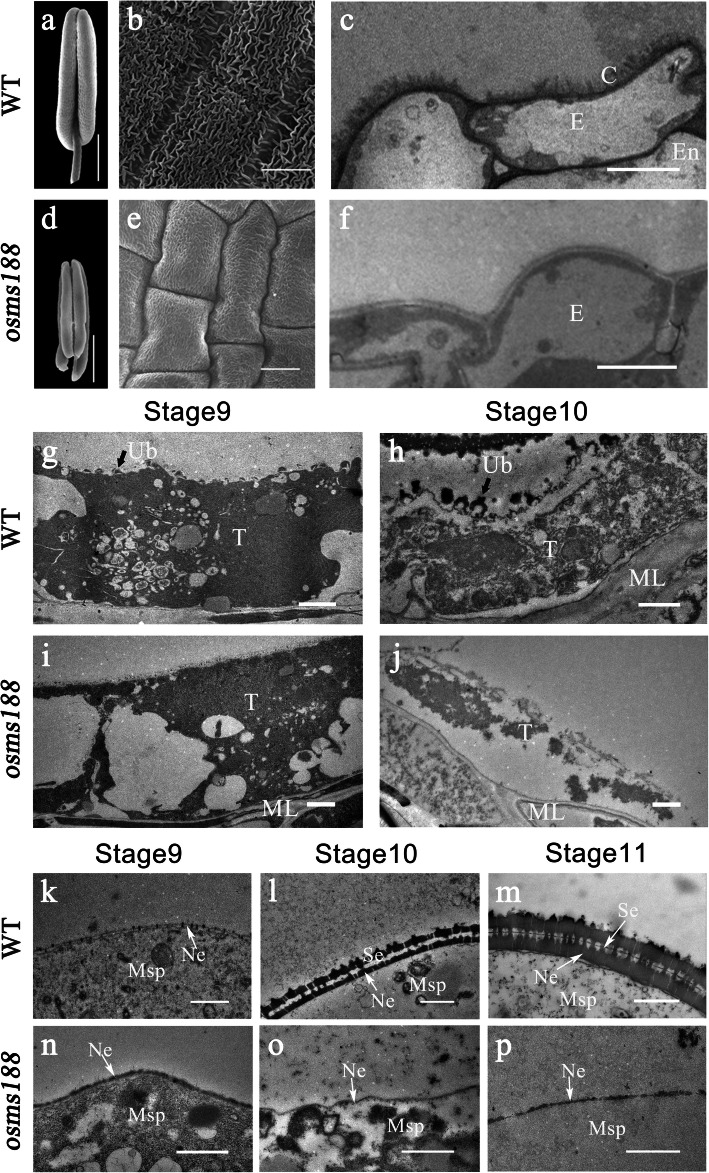
Fig. 2.
Electron micrographs of anthers and pollen grains of the WT and osms188 mutant. SEM observations showing the anther and pollen development of the WT (a-b) and osms188 (d-e). Anther morphology of the WT (a) and osms188 (d) and the epidermal surfaces of WT (b) and osms188 (e) anthers. TEM observations of tapetum development and sexine formation of the WT (c, g, h, k, l, m) and osms188 (f, i, j, n, o, p). Cuticle layer of the WT (c) and osms188 (f). The tapetal cells of the WT (g) and osms188 (i) at stage 9 and the tapetal cells of the WT (h) and osms188 (j) at stage 10. Pollen wall formation in the WT (k, l, m) and osms188 (n, o, p) during stages 9–11. C, cuticle; E, epidermis; En, endothecium; ML, middle layer; Msp, microspore; Ne, nexine; Se, sexine; T, tapetum; Ub, Ubisch body. Bars: (a, d) 500 μm; (b, e) 20 μm; (c, f) 5 μm; (g-l, o-p) 1 μm; (m, n) 0.5 μm

