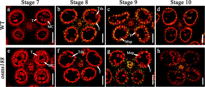
Fig. 3.
TUNEL analysis in the anthers of WT and osms188. The TUNEL signals are indicated with green fluorescence. The red fluorescence indicates nuclei stained with propidium iodide. At stage 7, no TUNEL signals were detected in the anthers of the WT (a) or osms188 (e). The TUNEL signals of the WT were normal at stage 8 (b), stage 9 (c) and stage 10 (d). Weak TUNEL signals were detected at stage 8 (f), stage 9 (g) and stage 10 (h) of the osms188 anthers. Bars = 50 μm. MC, meiotic cell; T, tapetum; Tds, tetrads; Msp, microspore

