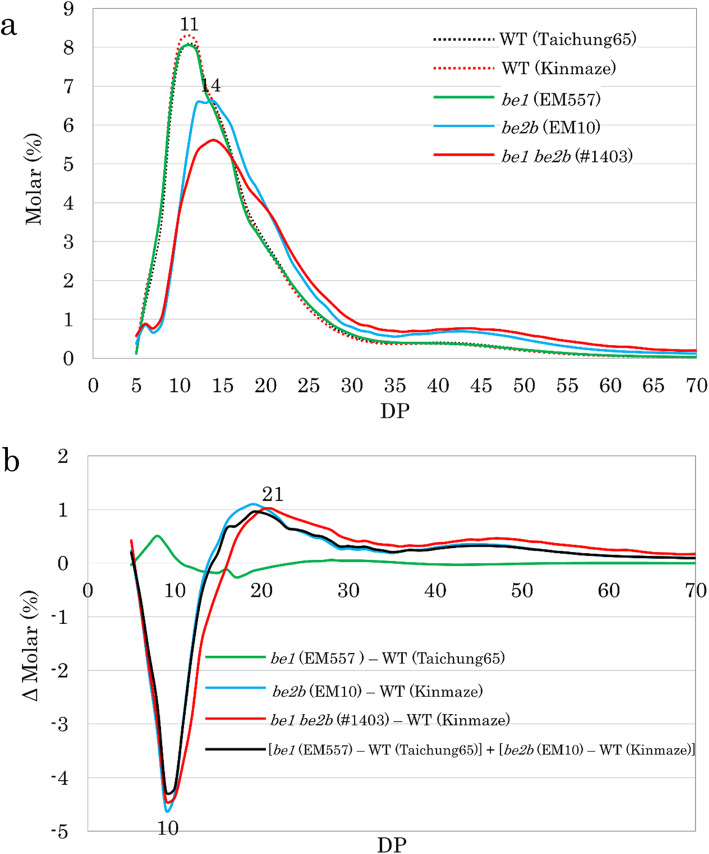
Fig. 5.
Chain length distribution of endosperm amylopectin analyzed by capillary electrophoresis. a Chain length distribution patterns of WT, be1, be2b, and be1 be2b. b Differences in the fine structure of amylopectin between the WT and mutant lines. Differences are shown as Δ Molar %, and the value was calculated by subtracting the pattern of WT from each mutant line, as indicated. Black line indicates a theoretical value calculated by adding the effects of the loss of BEI alone and BEIIb alone

