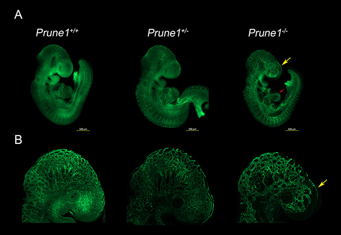
Figure 5.

Loss of Prune1 results in vascular defects with significant disruption of the cephalic vascular plexus. (A) Representative whole mount Pecam1 staining at E9.5 demonstrated reduced plexus branching and perturbed capillary sprouting within the cephalic region (frontonasal prominence and brain, yellow arrow) in the Prune1−/− embryos as compared with wild-type and heterozygous littermates. Moreover, Prune1−/− embryos displayed cardiac defects observed as a less intricate appearance of the endocardium when compared with littermate controls (red arrow) (B) Higher magnification (6.3X) images further highlight the disruption of the cephalic vascular plexus. A total of three Prune1+/+, 2 Prune1+/− and 5 Prune1−/− embryos were analyzed in two independent experiments.
