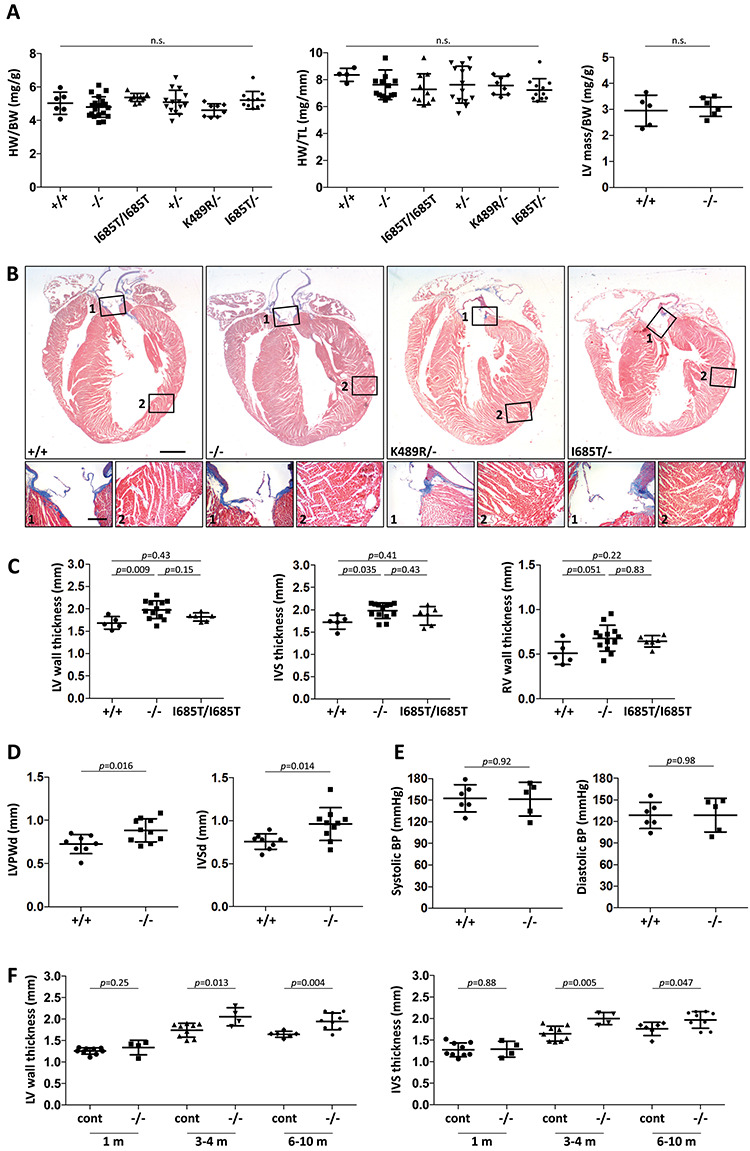
Figure 2.

Phenotypic evaluation of Tnni3k mutant mouse hearts. (A) Heart weight to body weight ratio, heart weight to tibia length and left ventricle mass to body weight ratio measurements for hearts of the indicated genotypes; all mice were 3–10 months old, and each dot represents a unique mouse. All pair-wise comparisons were statistically non-significant (n.s.). (B) Trichrome-stained long-axis histology sections of representative hearts from 7 month old mice fixed in diastole; the magnified regions (numbered) show positive collagen staining (blue) in the aortic valve (number 1 boxes) but little if any staining in the myocardium (number 2 boxes). All corresponding images are at the same magnification. Scale bar in larger images, 1 mm; scale bar for smaller images, 200 μ. (C) Measurements of left ventricular (LV) wall, interventricular septum (IVS), and right ventricle (RV) wall thicknesses in transverse sections taken at a cross-sectional plane through the papillary muscles. LV wall thickness was measured between the two papillary muscles, and IVS and RV thicknesses directly opposite. Representative images of sections are shown in Supplementary Material, Figure S1A. Analysis of additional genotypes (+/−, K489R/−, I685T/−) is shown in Supplementary Material, Figure S1B. (D) Measurement of LV posterior wall (LVPW) and interventricular septal (IVS) diastolic diameters in live mice by echocardiography. (E) Tail-cuff measurement of systolic and diastolic blood pressure in non-anesthetized mice. (F) LV and IVS thickness in control (+/+ and +/− combined) and null (−/−) mice at different ages as measured in transverse sections.
