Fig. 1.

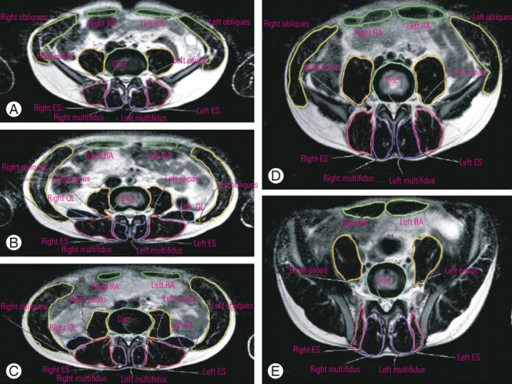
Measurements of the cross-sectional areas of trunk muscles on axial T2-weighted images at the (A) L1–L2, (B) L2–L3, (C) L3–L4, (D) L4–L5, and (E) L5–S1 lumbar disc levels. RA, rectus abdominis; ES, erector spinae; QL, quadratus lumborum.

Measurements of the cross-sectional areas of trunk muscles on axial T2-weighted images at the (A) L1–L2, (B) L2–L3, (C) L3–L4, (D) L4–L5, and (E) L5–S1 lumbar disc levels. RA, rectus abdominis; ES, erector spinae; QL, quadratus lumborum.