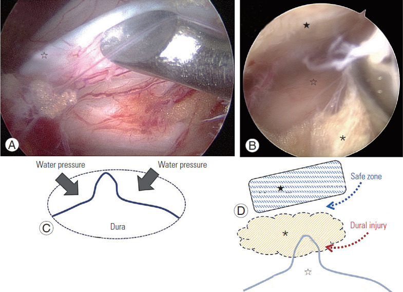
Fig. 2.

Central dural folding. (A, B) Intraoperative endoscopic image demonstrating central dural folding. There are three layers: ligamentum flavum (★), epidural fat layer (*), and central dural folding (☆). A curette is working on the epidural fat. (C) Effect of water pressure forming the central dural folding. (D) A safe zone for crossing the midline over central dural folding. Insertion of sharp instruments should be avoided under the epidural fat without caution of central dural folding.
