
Fig. 4.
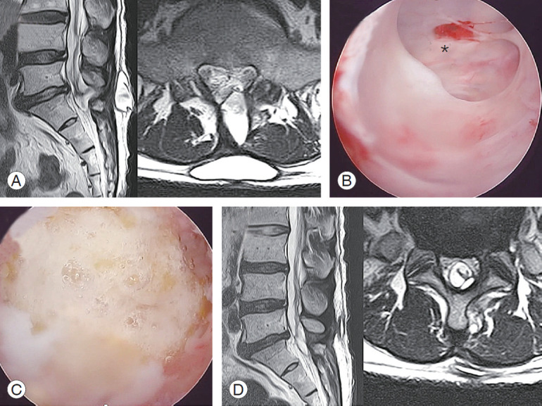
Pseudomeningocele. (A) A huge pseudomeningocele formed at the subcutaneous layer connecting to the deeper inside of the spinal canal at postoperative week 8 MRI. (B) Revision using biportal endoscopic spinal surgery into the pocket for debridement and to eliminate the pseudomembrane and trace the tract into the remnant orifice (*) of the dura. It is already healed and left as a crescent shape. (C) TachoSil is patched at the orifice. (D) Postrevision 4-week MRI shows a successfully resolved state. MRI, magnetic resonance imaging.