
Fig. 1.
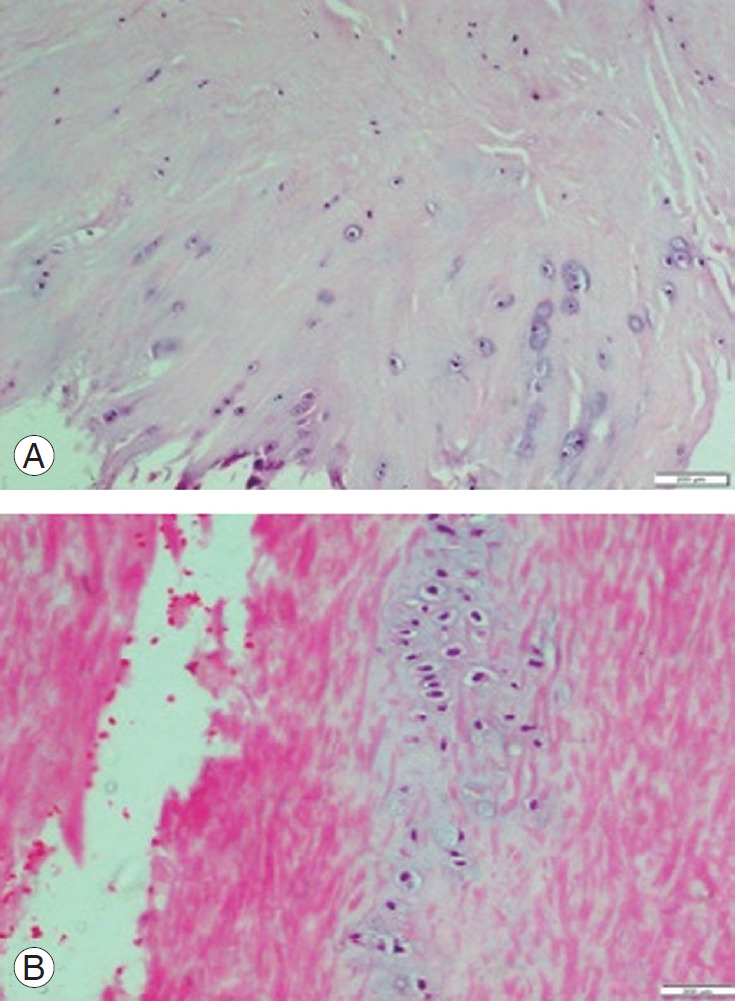
(A) HE stained section of ligamentum flavum in patient with degenerative lumbar canal stenosis showing areas of chondroid metaplasia at bony attachment. Chondrocytes arranged in pairs or in chains or clusters (H&E, ×100). (B) HE stained section of ligamentum flavum of a patient with disc herniation showing areas of chondroid metaplasia in the central region (H&E, ×200). HE, hematoxylin and eosin.