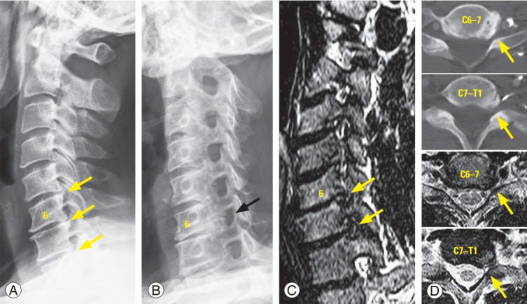
Fig. 1.

A 53-year-old male patient’s radiographs of cervical spine. He complained severe left-side scapular medial border pain and weakness of left finger extension. Intervertebral disc space narrowing and posterior ostephytes were observed in multiple levels (A, yellow arrows) and large bony spur and foraminal stenosis were well presented at C6–7 segment of oblique X-ray (B, black arrow). The C6–7 and C7–T1 foraminal stenoses seems to be more clear in oblique coronal images of magnetic resonance imaging (C) than in axial computed tomography scans or magnetic resonance images (D).
