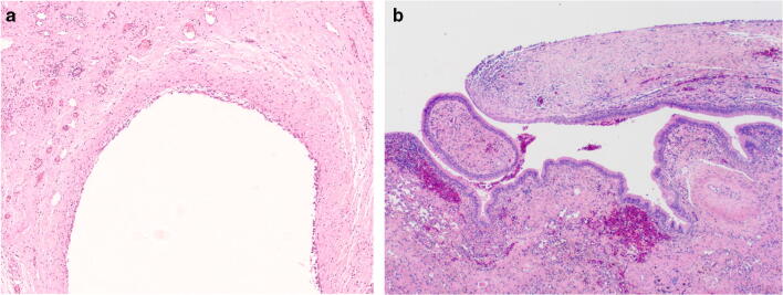
Fig. 2.
a Intermediate power view of the cystic space exhibiting a simple cuboidal epithelial lining and fibrotic wall associated with vascular congestion. b Intermediate power image of the fibrous cyst wall (right) transitioning with ciliated respiratory epithelium (left) (reprinted with permission from the European Respiratory Journal)

