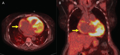
Figure 3: 18F-Fluorodeoxyglucose PET Imaging.

A: Axial and B: coronal PET images show 18F-fluorodeoxyglucose uptake in the atrium (yellow arrows) in a patient with AF.

A: Axial and B: coronal PET images show 18F-fluorodeoxyglucose uptake in the atrium (yellow arrows) in a patient with AF.