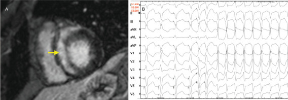
Figure 4: Late Gadolinium Enhancement on Cardiac MRI and Electrophysiological Testing.

A: Late gadolinium enhancement on cardiac MRI shows substantial enhancement involving the ventricular septum (yellow arrow). B: On electrophysiological testing, the patient exhibited easily inducible monomorphic ventricular tachycardia.
