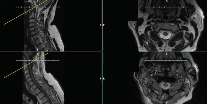
Fig. 4.
The discrepancy between axial and sagittal cuts on magnetic resonance imaging of the cervical spine. Top left and bottom left show a T2- and T1-weighted sagittal cuts, respectively, demonstrating 2 different cervical spine levels. However, the top right and bottom right images show T2- and T1-weighted axial cuts, respectively, that both are at the same level (at the base of C2).

