
Fig. 4.
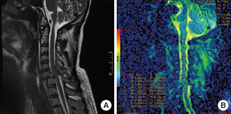
The diffusion tensor imaging of a patient with C6/C7 spinal cord injury. (A) Sagittal image of the T2-weighted image of the cervical spine. (B) Diffusion tensor imaging sagittal section. Reprinted from Czyz et al. J Spinal Study Surg. 2017;1:25-28, under the terms of Open Access [30].