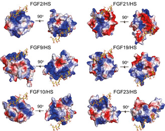
Figure 3. Heparin binding sites of FGF21 differ from that of paracrine‐acting and other endocrine‐acting FGFs.

Electrostatic potential surface of different FGFs for representing the (HS) binding sites. HS is placed by superimposition of FGF21core, FGF9 (PDB ID: 1IHK), FGF10 (PDB ID: 1NUN), FGF19 (PDB ID: 2P23), and FGF23 (PDB ID: 2P39) onto the FGF2/FGFR1c/HS ternary complex structure (PDB ID: 1FQ9). HS is shown as a stick representation. The positive and negative charges on the protein surface are colored blue and red, respectively.
