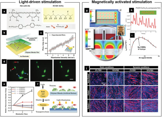
Figure 8.

Light‐driven /Magnetically activated piezoelectric stimulation of cells in vitro. a) Molecular structures of azobenzene polymer P(8‐AZO‐10) and its conversion of trans and cis isomers upon photoirradiation. b) Layout of a ferroelectric polymer arrays as photodetector, consisting of ITO coated PET, 10‐µm‐thick P(VDF‐TrFE)/P(8‐AZO‐10) blends, and Au electrode. c) Profile of piezoresponse amplitude as a function of light intensity. d) Calcium‐ion imaging of PC12 cells cultured on the blend membrane in response to the white LED stimulation. e) The change of fluorescence intensity for the cells seeded on the blend membrane or not. f) Scheme of the signal transduction from the photoreceptor cells (left) or the photodetector to the neuron cells (right). Reproduced with permission.[ 84 ] Copyright 2016, Wiley‐VCH. g) Illustration of simulated magnetic field and the culture setup for myoblast. h) Measurement mechanical strain induced by the applied dynamic magnetic field with the change of time. i) magnetoelectric response of the CFO/P(VDF‐TrFE) composite film for a DC field from 0 to 5000 Oe at a constant Hac of 1 Oe. j) Immunofluorescent staining of myosin heavy chain after 5 days of C2C12 cells differentiation on the different samples. Reproduced with permission.[ 85 ] Copyright 2020, American Chemical Society.
