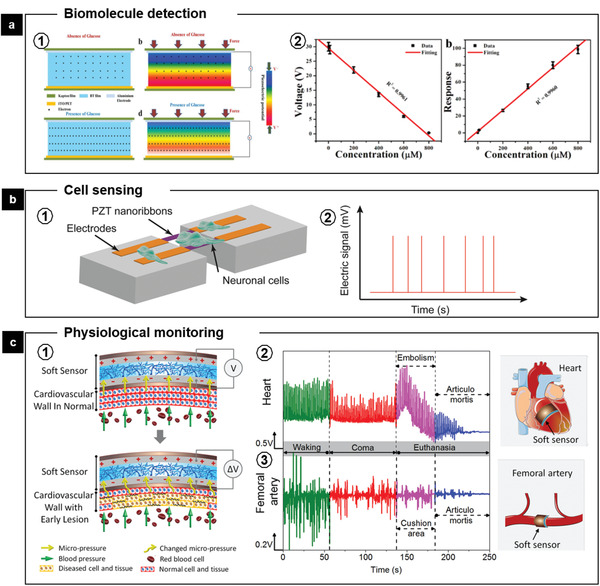
Figure 12.

FEMs‐mediated biosensing. a1) Schematic representation of BTO film‐based NG during active biosensing of glucose. a2) Relationship between the piezoelectric output of NG and the concentration of glucose and dependence of the response on the concentration of glucose. Reproduced with permission.[ 93 ] Copyright 2017, Elsevier. b1) Schematic of the piezoelectric PZT nanoribbon device with cultured neuronal cells. b2) Illustration of the response of piezoelectric nanoribbons to cellular deformations evoked by an applied membrane voltage.[ 94 ] c1) Schematic of the working principle of the sensor for sensing micropressure changes caused by the early lesion of the cardiovascular wall, which usually led to the changing on the mechanical characteristics or physiological structure of the cardiovascular wall. Output piezoelectric signals of the sensors implanted on c2) heart or c3) femoral artery induced by the cardiovascular elasticity changes of pig at different physiological states. Adapted with permission.[ 98 ] Copyright (2019) American Chemical Society.
