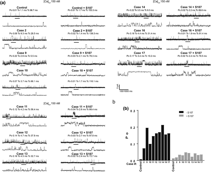
Figure 2. Increased calcium leak in single RYR1-RM channels reconstituted in planar lipid bilayer.
SR microsomes containing single RyR1 channels isolated from individuals with RYR1-RM fused with planar lipid bilayer. Channel opening events are recorded as an upward deflection. Area on upper graph is expanded as the lower graph. Po = opening probability, To = time open, Tc = time closed. (a) Single-channel recordings of RyR1 from muscle lysates that were either treated or untreated with S107 (1.0 μM) from control and RYR1-RM affected individuals. Recordings were performed at 150 nM Ca2+. (b), Bar graph summarizing single-channel Po. N=3 per group. Limited availability of skeletal muscle precluded analyses in Cases 3–8 and 15.

