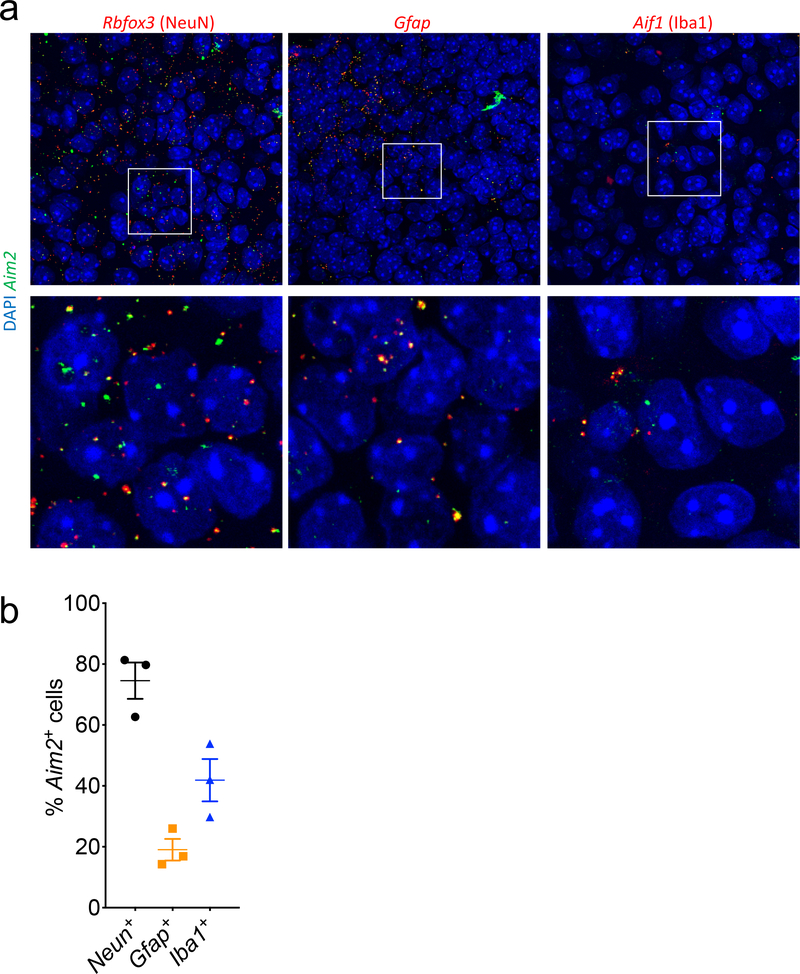
Extended Data Figure 9. Aim2 is expressed by neurons, astrocytes, and microglia in the developing brain.
Brains from p5 WT mice (n=3; from 1 experiment) were evaluated for expression of Aim2 using RNA scope. (a) Images showing co-expression of Aim2 (green) and CNS cell-specific genes Rbfox3: NeuN (red), Gfap: GFAP (red), and Aif1: Iba1 (red) in the hippocampus. (b) Quantification showing percentage of CNS cells in 40X images that are positive for Aim2. Error bars depict mean ± s.e.m. n values refer to biological replicates.

