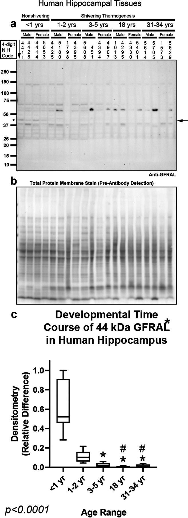
Fig. 7.

Developmental time course of GFRAL in the human hippocampus. a Representative blot of relative GFRAL levels in the hippocampus in infants (n = 8/group), toddlers (n = 8/group), preschoolers (n = 7/group), adolescents (n = 8/group), and adults (n = 8/group). b Membrane stain shows total protein loading across subjects. c Normalized densitometry values were analyzed by Kruskal-Wallis 1-way ANOVA. * indicates post hoc significance vs. infants. # indicates post hoc significance vs. toddlers. Data were significant at p < 0.05. Box plots show minimum, maximum, IQR, and median. The asterisk above GFRAL denotes that the band analyzed by densitometry matches the predicted kDa
