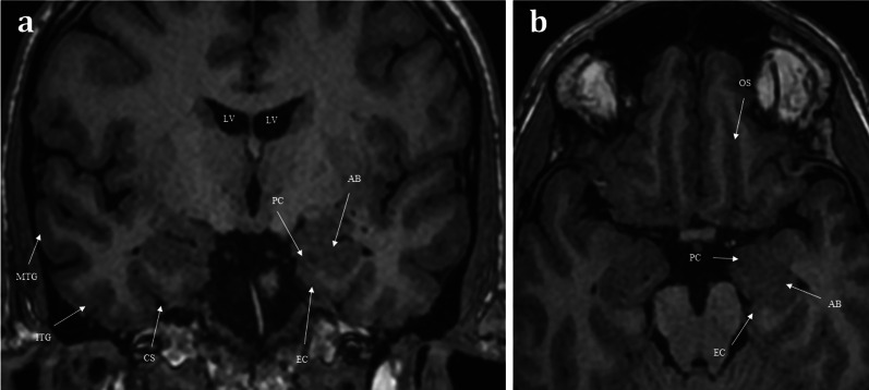
Fig. 3.
Coronal T1 weighted image taken at the level of the lateral ventricles (LV) and foramina of Monroe (a), and axial T1 weighted image of the inferior frontal lobe and mesial temporal lobes (b). The olfactory tracts (not pictured) run within the olfactory sulci (OS) and transmit second-order neurones to the central olfactory regions. These are housed within the mesial temporal lobe and include the parahippocampus, the entorhinal cortex (EC), the piriform cortex (PC), and the amygdaloid body (AB). Note the other sulcal landmarks including the collateral sulcus (CS) which bounds the entorhinal cortex inferiorly, as well as the inferior temporal gyrus (ITG), and the middle temporal gyrus (MTG)

