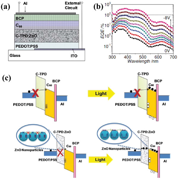
Figure 8.

a) Schematic device structure of the photodetector with ZnO nanoparticles mixed in the C‐TPD buffer layer. b) EQE spectra of the photodetector under reverse bias from 0 to −8 V with a voltage step of 1 V. c) Energy level diagram of the reverse‐biased photodetector in dark and under illumination. Reproduced with permission.[ 79 ] Copyright 2014, Wiley‐VCH.
