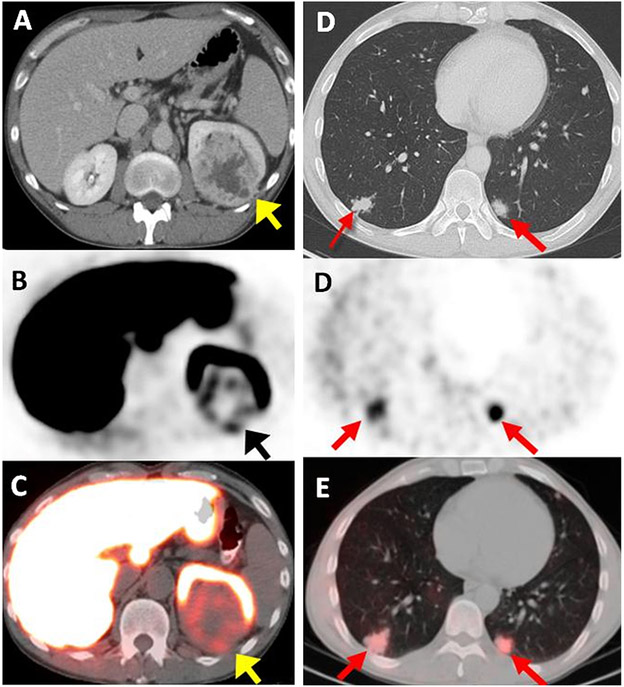
Fig. 2.
A 44-year-old male with left-sided ccRCC detected on contrast enhanced CT (arrow) (A). Axial 18F-VM4-037 PET (B) and PET/CT (C) show uptake within the left kidney lesion (arrow) with an SUVmean of 2.45. Axial CT shows bilateral metastases in lung bases (SUV = 2.22 in the right lower lobe lesion with histology confirmation of poorly differentiated ccRCC) (arrows) (D). Axial 18F-VM4-037 PET/CT (E) show uptake of 18F-VM4-037 both lesions (arrows).

