Fig. 5.
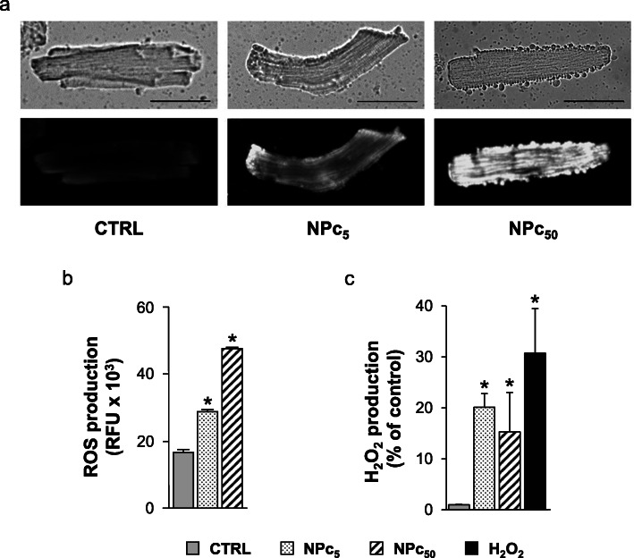
ROS production in rat ventricular cardiomyocytes exposed to Co3O4-NPs. Intracellular ROS amount was measured in untreated (CTRL) or NP-treated cardiomyocytes at 5 μg/ml (NPc5) and 50 μg/ml (NPc50) after 1 h exposure at room temperature using different methodologies. a) Cardiomyocytes were stained with CellROX® Orange Reagent and visualized with fluorescence microscopy. A representative phase contrast (upper-side) and fluorescence (lower-side) microscopy images were shown. Scale bars were set at 50 μm. b) Reactive species (ROS/RNS) production detected using DCFDA assay were expressed as relative fluorescence units (RFU). *, p< 0.05 vs CTRL (one-way ANOVA; Bonferroni post-hoc test). c) Amount of H2O2 produced by cardiomyocytes was detected using Amplex red Hydrogen Peroxide/Peroxidase assay and expressed as RFU increment (%) respect to untreated control. As positive control, cardiomyocytes treated for 20 min with 0.003% H2O2 (0.88 μM) were also analyzed. *, p< 0.05 vs CTRL (Kruskal-Wallis test followed by U-Mann Whitney test)

